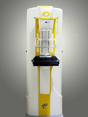
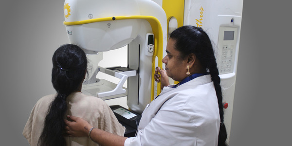
Breast cancer remains an important public health issue. It is the leading cause of cancer-related deaths in India and in recent years is emerging as the commonest female malignancy, overtaking cancer of the cervix. There are an estimated 1,00,000 -1 ,25,000 new breast cancer cases in India every year. Early detection of breast cancer plays a leading role in reducing mortality rates and improving the patient's' prognosis. The most frequently recommended screening method for early detection of this fatal disease is mammography
What is Mammography?
A mammogram is an x-ray picture of the breast and is used to check for breast cancer in women who have no signs or symptoms of the disease. It is a widely used imaging modality for breast cancer screening. A mammography exam, called a mammogram, aids in the early detection and diagnosis of various breast diseases in women.
When should Mammograms be done?
For most women, who are not at especially high risk of breast cancer, regular mammograms can start at age 50. Or, to be cautious, a woman can get one mammogram earlier (around age 45) and then if it is normal, wait until she is 50 for her next mammogram.
- For a woman at high risk of breast cancer because of her family history or environmental exposures, regular screening before age 40, is highly recommended.
- Women who are carriers of the BRCA genetic mutation are recommended to begin yearly mammograms between ages 25-30, since this mutation puts them at much higher risk of getting breast cancer.
Commonly Used Mammography Techniques
Let’s have a look at the widely used mammography procedures.
Screen Film Mammography
Screen-film mammography has long been considered as a “gold standard” for breast cancer screening. In addition to its ability to provide adequate visualization of soft tissue abnormalities, its particular strength is the ability to depict subtle calcifications. Screen-film mammography is a powerful tool for initial detection and in the subsequent follow-up of suspicious lesions.
Digital Mammography
 The term “digital mammography” is used for any technology which employs a single or multiple detector assembly to capture an electronic image of the x-rays transmitted through the breast that can be displayed, stored, and communicated electronically.
Digital breast tomosynthesis is an imaging technique designed to eliminate the pitfalls of overlapping tissue. It has the potential to lower recall rates on screening mammography and reduce false negative examinations due to dense breast tissue. Tomosynthesis produces tomographic “slices” of an entire tissue volume, similar to a CT scan, using a single acquisition.
Common use of the Procedure
Mammograms are used both as a screening tool to detect early breast cancer in women experiencing no symptoms and to detect and diagnose breast disease in women experiencing symptoms such as a lump, pain, skin dimpling or nipple discharge.
What are Screening Mammograms?
Conceptually, screening mammography implies a mammographic examination performed on a woman who has neither a palpable mass nor symptoms of breast cancer. The components of a breast screening evaluation depend on patient age and other factors, such as medical and family history, and can include breast awareness (i.e., patient familiarity with her breasts), physical examination, risk assessment, screening mammography, and, in selected cases, screening MRI.
Also Read: Dense Breasts & Cancer Risks – End the Confusion!
Difference between a Screening and a Diagnostic Mammogram
A diagnostic breast evaluation differs from breast screening in that it is used to evaluate an existing problem. Diagnostic mammography is commonly used to identify possible breast cancers in women who present with signs or symptoms of the disease. These signs or symptoms may include a palpable breast lump, nipple discharge or retraction, and breast dimpling or other skin changes. A diagnostic mammographic examination usually consists of standard screening views and additional views using spot compression and/or magnification of a specific area. Although mammography may be sufficient to evaluate the clinical finding, additional imaging with ultrasound, ductography, or other imaging techniques may also be done to confirm the diagnosis.
Limitations of Mammogram
False negatives
This means everything may look normal, but cancer is actually present. False negatives don't happen often. Younger women are more likely to have a false negative mammogram than are older women. The dense breasts of younger women make breast cancers harder to find in mammograms.
False positives
This is when the mammogram results look like cancer is present, even though it is not. False positives are more common in younger women, women who have had breast biopsies, women with a family history of breast cancer, and women who are taking estrogen, such as menopausal hormone therapy.
Mammograms (as well as dental x-rays and other routine x-rays) use very small doses of radiation. The risk of any harm is very slight, but repeated x-rays could cause cancer. The benefits however outweigh the risk of succumbing to cancer.
