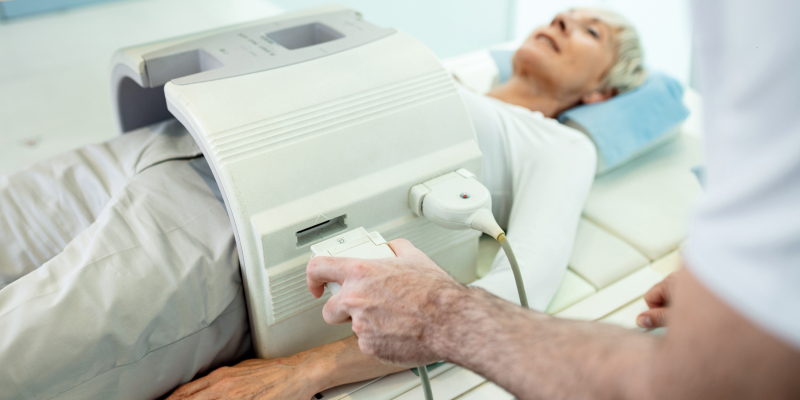
Ever wondered about the mechanics of an Abdomen MRI? Although it might appear complex, it's simpler than you think. This article will clarify the technical aspects and highlight why it's an essential tool for diagnosing a wide range of health conditions. It's normal to feel a bit anxious about an Abdomen MRI, and that's perfectly fine. The best scan center in Chennai is here to assist you. We'll explain the technology that powers it, prepare you for what to expect during the procedure, and help you understand your results. Are you ready for some clarity? Let's get started.
Understanding the Abdomen MRI
In your journey to grasp the details of Abdomen MRI, it is essential to understand the technology that drives it. This understanding will clarify the process, showing you how magnetic fields and radio waves collaborate to produce detailed images of your abdomen. This technical understanding could make the procedure seem less intimidating. Let's talk about the technology that powers Abdomen MRI.
The Technology Behind Abdomen MRI
The technology that powers an Abdomen MRI employs sophisticated machinery, magnetic fields, and radio waves to produce detailed images of your abdomen. It provides a clear vision of your body's structures, unseen with other imaging techniques like X-rays, ultrasounds, or CT scans.
In certain situations, a contrast material might be involved in the abdominal MRI scan to enhance the visibility of specific structures in the images. This enhancement assists in:
- Monitoring blood flow
- Detecting certain types of tumors
- Highlighting areas of inflammation or infection
The MRI process leverages radio waves and a robust magnetic field, eliminating the need for X-rays and thus, radiation. Some MRI exams might necessitate an injection of a contrast material named gadolinium.
The shift from traditional PACS to web-based imaging systems is underway, simplifying access to various imaging techniques. These systems blend access to AI and advanced imaging tools, and the data interfaces with Electronic Medical Records (EMRs) almost flawlessly.
Off-site cloud storage is increasingly becoming the preferred choice for storing images and patient data, a trend spearheaded by companies specializing in data centres. The latest generation MRI scanner boasts technical performance features that outshine existing whole-body MRI systems.
Artificial Intelligence (AI) is steadily becoming a crucial component of medical imaging, enhancing the skills of technicians and radiologists. Here are some of the advantages:
- Enhances efficiency
- Amplifies image quality
- Helps in reducing radiation dosage
- Streamlines data organisation, making it simpler to retrieve past data, auto-complete structured reports, and automate measurements.
Now that you have a good understanding of the technology, let's move on to its purpose.
The Purpose of an Abdomen MRI
Are you thinking about an abdomen MRI? This potent diagnostic tool assists doctors in identifying a variety of conditions. It has the capability to expose the inner workings of your body, from liver diseases to digestive disorders. With this information, you, along with your healthcare team, can strategize the most effective course of action. Let's discuss the typical conditions that an abdomen MRI can diagnose.
Common Conditions Diagnosed with Abdomen MRI
An Abdomen MRI serves as a versatile tool that diagnoses a variety of conditions. For example, it identifies liver conditions like cirrhosis and abnormalities in the bile ducts and pancreas. It also proves effective in diagnosing conditions of the gastrointestinal tract, such as Crohn's disease and ulcerative colitis, which fall under the category of inflammatory bowel diseases.
Moreover, an Abdomen MRI assists in diagnosing abnormal or inflamed blood vessels, a condition known as vasculitis. It frequently diagnoses conditions that cause severe abdominal pain, like gastroenteritis and appendicitis.
Specifically, an MRI diagnoses emergent gastrointestinal diseases quickly, such as:
- Choledocholithiasis
- Acute cholecystitis
- Acute pancreatitis
- Bowel inflammation in the setting of inflammatory bowel disease
Having understood the conditions that can be diagnosed with an Abdomen MRI, let's now delve into the procedure of an Abdomen MRI to understand how it aids in these diagnoses.
Also Read: MRI Scan- Definition & Methodology
The Procedure of an Abdomen MRI
Understanding the technical aspects of an abdomen MRI involves knowing the procedure and the preparations required. This knowledge includes your dietary guidelines, dress code, and the use of contrast agents during the procedure. You will also learn about the sensations you might experience. Let's look at these aspects in more detail.
Preparation for an Abdomen MRI
To achieve a clear and successful Abdomen MRI, follow these steps- Fast for about 4 hours before the exam and take your regular medications with a bit of water. If your scan requires contrast material, the physician might use gadolinium to improve the MRI images. Allergic reactions to gadolinium are rare, but discuss any potential allergies with your physician before receiving it.
For scans requiring contrast material, adhere to your doctor's instructions about when to stop eating and drinking. If the physician needs images of your gastrointestinal tract, you'll need to prepare your bowel using laxatives or enemas, and fast for 4 to 6 hours before the exam.
In terms of attire and personal items, wear comfortable clothing and prepare to change into a hospital gown when you arrive at the radiology facility. Leave all personal items, especially metal objects like jewellery, at home to prevent any interference with the MRI machine. If you have an artificial cardiac pacemaker or any implanted metal in your body, inform your physician as they might suggest alternative radiological examinations like a CT scan.
If you feel anxious in confined spaces, tell your physician, who may provide medication to help you relax. However, if you receive such medication, arrange for a ride home after the exam. If you're preparing for a pelvis MRI, drink plenty of fluids before the exam to ensure a full bladder.
With these preparations, you're ready for your Abdomen MRI. Next, we'll discuss what you can expect during the procedure.
During the Abdomen MRI
The Abdomen MRI procedure starts with you lying face up on the MRI table. The technologist slides the table into a large, tunnel-shaped magnetic scanner. The scanner makes a series of humming, clicking, and knocking sounds, but you won't feel anything. In some scenarios, the MRI technologist might inject a contrast agent, or dye, into a vein in your arm or hand. This injection helps capture more detailed images and takes about 1 to 2 minutes. After the injection, the technologist performs more MRI scans.
For certain MRI tests, you might receive a medication like glucagon to slow down your bowel movements. The technologist instructs you on when and how to breathe during the scan. It's crucial to stay as still as possible because the MRI machines react extremely to movement. At times, the technologist might ask you to hold your breath while capturing images. The technologist remains in contact with you during the scan and provides you with an emergency device. You can use this device to alert them if you need help.
The procedure doesn't cause any pain, but you might feel enclosed. However, the use of wide-bore MRI scanners has significantly reduced this feeling for most patients. Now that you understand the procedure, let's discuss what happens after the scan.
Interpreting the Results of an Abdomen MRI
If you've just completed an abdomen MRI, you might find the report filled with medical terms and findings that seem challenging to understand. Fear not, this section aims to simplify your report for easy comprehension. So, let's begin the process of understanding your Abdomen MRI report.
Understanding Your Abdomen MRI Report
Understanding your Abdomen MRI report is vital for your active involvement in your healthcare. The report divides into several sections, each delivering crucial insights into your abdominal health:
- 'Type of exam': This section states the date and type of exam.
- 'History/Reason for exam': This section details your symptoms and the test's reason.
- 'Comparison/Priors': This section enables the radiologist to compare the new exam with any previous ones you've had.
- 'Technique': This part describes the exam process, including the use of contrast or not.
- 'Findings': This section, the most crucial part, summarises the findings and suggests potential causes.
Your Abdomen MRI offers a detailed view of various parts of your abdomen, including:
- Organs: liver, spleen, and pancreas
- Parts of the bowel
- Lymph nodes
- Blood vessels
It can identify cancers, inflammation, and provide a better visualisation of the blood supply of organs and blood vessels.
In a normal scenario, your organs and blood vessels should appear normal in size, shape, and location. There should be no abnormal growths or blockage in the ducts draining the liver, gallbladder, or pancreas. However, if any organ is too large, too small, or in the wrong place, or if there are areas of scarring or injury, or signs of infection, it may indicate an abnormality. Understanding this will allow you to better comprehend the results of your Abdomen MRI and take an active role in your healthcare decisions.
Empowering Knowledge for Healthier Decisions
An Abdomen MRI, a non-invasive, potent diagnostic tool, uses magnetic fields and radio waves to create a clear image of your internal organs. Understanding why doctors perform it and how to prepare can help alleviate any anxiety. Knowing your results empowers you to take an active part in your medical decisions. And, if you need to book a consultation with the best MRI scan centre in Chennai, get in touch with us at Anderson Diagnostics. As Benjamin Franklin wisely said, "An investment in knowledge pays the best interest." So, don't hesitate to expand your understanding or seek additional help from professionals.
Frequently Asked Questions
What is the purpose of abdominal MRI?
An abdominal MRI, a harmless procedure, uses magnetic and radio waves to generate detailed images of the abdominal area. Medical professionals use this procedure to examine soft tissues without bone interference. When a physical examination fails to diagnose a potential abdomen issue, they turn to an abdominal MRI. It helps detect conditions like gallstones, liver disease, tumors, and infections. It also helps find the root of abdominal discomfort or bloating, studies blood flow and vessels, and assesses the health of organs such as the liver, pancreas, and kidneys.
How does an abdomen MRI work?
This technique lets medical experts search for any abnormalities in the tissues and organs without surgical intervention. The MRI machine can produce 3D images for viewing from different angles. One more benefit of an MRI is its lack of radiation, making it safer than a CT scan.
What are the risks and side effects of an abdomen MRI?
Typically, an abdominal MRI is safe, presenting no known side effects from its use of radio waves and magnetism. However, it's worth noting that the MRI machine's magnets may attract body metals. Hence, if a person has metal implants or injury fragments, they must inform the doctor. Some people might experience discomfort due to the machine's confined space, potentially triggering claustrophobia. In rare instances, the contrast dye used in MRIs could lead to allergic reactions or kidney damage. Even less common side effects might be nausea, vomiting, constipation, diarrhea, skin rashes, headaches, or a flushed complexion.
How long does an abdomen MRI take?
Typically, an abdominal MRI scan takes between 30 to 60 minutes. But, the duration can vary based on factors like the MRI machine type, the specific imaging area, and the complexity of the case. Using a contrast dye during the procedure may extend the time, as it requires capturing more images. Overall, an MRI scan process can range between 15 to 90 minutes. Each image from the scan generally takes about 3 to 4 minutes to complete. Often, doctors require multiple images to aid in diagnosis.
What does it feel like to have an abdomen MRI?
An abdominal MRI is a straightforward, pain-free procedure. You may find the table a bit hard and cold, but a blanket or pillow can increase your comfort. Expect loud thumping and humming noises from the machine during operation. If you're not a fan of tight spaces or prone to nervousness, a prescription for a sedative or antianxiety medication can help maintain your calmness. Remaining still during the scan is essential since any movement can blur the images, potentially causing errors.
When will I get the results of my abdomen MRI?
Typically, MRI scan results take around one to two weeks. But if the case is urgent, they deliver the results faster. A radiologist looks at the images and sends a report to your doctor. Then, your doctor arranges a meeting with you to talk about the results. Keep in mind, these timelines can vary. It depends on the hospital or clinic you visit and how serious your medical situation is.
How accurate is an abdomen MRI?
An abdominal MRI's effectiveness can fluctuate based on the specific health issue under observation. For instance, early results suggest that MRI may help to assess diverticulitis, correctly diagnosing the condition in 86% to 94% of cases and accurately dismissing it in 88% to 92% of cases. Another study found MRI's capacity to accurately diagnose and dismiss acute appendicitis at 97% and 93% respectively. However, using this method to visualize the function of the small and large bowel (intestines) becomes more challenging. It's important to note that while MRI surpasses a CT scan in precision for some health issues, it falls short for others.
