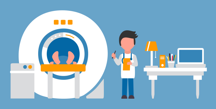

CT Scanner throws a series of narrow beams on the body while moving in an arc. It is not an X-Ray machine where just one radiation beam is sent. Here you can get a more elaborate picture in comparison to the X-ray image. The detector inside the scanner can see hundreds of levels of density. The tissues in the solid organs can be seen. The data is later transmitted to a computer, and a 3 D image is built. A CT scan machine looks like a huge doughnut with a tipped side. The patient is made to lie on the platform, and the patient is made to move into the hole in the machine. Then the X-Ray tube is mounted around the edges on a movable ring. There are X-ray detectors on the opposite side of the tube. Once the motor is on, the tube and the detector revolves around the body. For every full revolution, a horizontal slice of the body is scanned. The platform is moved further to continue the process for the next slice.
Cardiac CT scan is a non-invasive procedure where the X-ray images of the heart anatomy, aorta, coronary circulation, arteries and pulmonary veins. Each part of the heart is pictured, and the computer processes it into a 3 d image. It is used to evaluate conditions like calcium buildup, heart valve problem, aortic dissection, aortic aneurysm, pericardial disease, heart function problem, pulmonary embolism and peripheral vascular atherosclerosis.
CT Scan is performed to evaluate a tumour or lesions, injuries, structural anomalies, intracranial bleeding and other conditions of the brain. It is used to assess the treatment for a brain tumour and detect clots responsible for strokes. CT Scan of the brain is performed with an X-Ray and other examinations do not provide accurate results.
