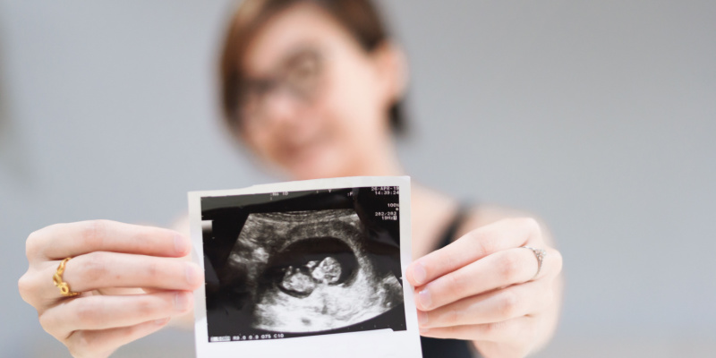
There are numerous turning points in one's life. One of the most exciting parts of being pregnant is finding out that you will be a parent and seeing images of your unborn child for the first time through an ultrasound. In addition to a positive pregnancy test and severe morning sickness, the first ultrasound, typically occurring at around 12 weeks, proves that you are indeed carrying a baby. And well, if you are looking for a truly reliable diagnostic centre in Chennai for this purpose, you can never go wrong with the Anderson Diagnostics scan centre.
Despite all the awe you'll feel seeing your unborn child for the first time, it's important to remember that your 12-week scan is essential to your pregnancy. It can give you and your doctor some valuable insights. If you're anxious or want to know more, you'll find all the information below.
Why 12 weeks?
Sonography, also known as ultrasonography, is an imaging technique that uses the energy from sound waves to create images of the internal anatomy.
The scan performed at 12 weeks of pregnancy, can provide valuable information about when the baby is expected to be born. This is helpful if you have irregular periods or can't remember the date of your last period.
Points to Note When You Visit Your Nearest Scan Centre in Chennai
Your 12-week ultrasound will go more smoothly if you take the following precautions:
- An ultrasound will yield more precise results if you hydrate yourself by drinking 2-3 glasses of water allowing the waves to travel smoothly on the fluid.
- Dress comfortably in loose-fitting clothes that won't restrict your motion. The scan may require you to undress from the waist down, so wear something that can be quickly taken off.
- The 12-week ultrasound can be an exciting and nervous experience. So, keeping calm is essential.
It's important to understand that there are two primary kinds of scans: transabdominal and transvaginal. Both involve a scan across the abdomen, but one also goes across the vagina.
In most cases, a transabdominal ultrasound, which sends sound waves through the belly, will be performed by your doctor around the 12-week mark. A transvaginal ultrasound may get clearer or more in-depth images in certain situations. It takes about twenty to thirty minutes to finish a scan.
Scan of the Abdomen with a Transducer
Your abdomen will be exposed from your ribs to your hips as you lie on an exam table during a transabdominal ultrasound, which can be done in a hospital or doctor's office. Your doctor may request that you come to the meeting with a full bladder to facilitate a clear view of the uterus.
At the start of the exam, your doctor will apply a gel that will help the ultrasound waves penetrate your skin. As a result, the ultrasound images will be of higher quality.
A portable ultrasound transducer will then be gently moved back and forth throughout your abdomen by your healthcare provider. This shouldn't hurt, though you might feel pressure depending on your position.
They might linger over your stomach for a moment while they take pictures or measurements. The baby and your uterus will be measured at various stages of pregnancy. Your baby's heartbeat could be recorded for a brief period.
While you don't need to empty your bladder before a transabdominal ultrasound, you will be asked to do so for a transvaginal scan. The technician may ask you to change into a hospital gown so that they can work on your lower body.
To get a better look at your uterus, your doctor will insert a wand-shaped transducer into your vagina while you lie on your back with your knees raised or your feet in stirrups.
Like the one used in a transabdominal scan, the transducer sends sound waves that reflect off internal organs and then send that information back to the screen. Although the transvaginal and transabdominal 12-week scans may sound unpleasant, they are painless.
The good news is that most ultrasounds at the 12-week mark are performed transabdominally.
Also Read: COVID-19 Travel Testing: The Ultimate Guide
Primary Lookouts of the Sonographer
Your sonographer's primary focus at the 12-week scan will be on the uterus, placenta, and fallopian tubes. A tilted uterus or placenta previa, for example, can cause complications during pregnancy and labour, and these conditions will be screened for the first time during your 20-week checkup.
An ultrasound at 12 weeks is your best bet for detecting these conditions, rare as they may be. When you visit the doctor between weeks 10 and 12, they will likely check for the following:
- Size of the baby: The length of your child from head to bottom will be measured at the 12-week ultrasound. The baby will be about 2.5 inches in length at this point. It's about the size of 2.5 paper clips!
- Placenta placement: The placenta is a crucial organ that supplies nutrients to the growing foetus. The sonographer will ensure the baby is resting in the best possible position for the baby's development and the mother's health.
- Baby’s heartbeat: One of the most exciting parts of the 12-week ultrasound is hearing your baby's heartbeat for the first time. For many moms and dads, this is a moment of great joy.
- Foetus’s development- The quantity of amniotic fluid serves to cushion and nourish the developing infant. A sufficient amount of amniotic fluid will protect the developing foetus, and the sonographer will verify this.
- Estimated delivery date: At the 12-week ultrasound, your due date can be estimated with reasonable precision. Though "accuracy" is somewhat vague, you would still get an approximate date.
- Testing for nuchal translucency: Conditions like Down and Edwards syndrome can be detected by examining the baby's nuchal translucency. Such tests are rarely performed unless the baby is thought to have a slightly increased risk of developing one of these conditions.
- Possibility of twins: The 12-week ultrasound is typically the first chance a pregnant woman has to learn whether she is carrying a single child, multiples, or more.
What if there's a problem?
In most cases, your 12-week ultrasound will go smoothly, and you can take home some stunning scans of your unborn child. The sonographer, however, may suggest additional testing if they discover anything of concern. Safe prenatal testing options include amniocentesis and chorionic villus sampling (CVS).
Talking to your doctor about any concerns you may have after receiving abnormal results from your 12-week ultrasound is essential.
Just a quick wrap!
Yes, Now that you have your 12-week ultrasound results and a heartbeat ringtone (no judgement here), you can begin preparing for the birth. It is never easy for first-time parents. You learn something new almost every day. But the arrival of your baby makes it all worth it. With our expert professionals at Anderson Diagnostic’s scan centre, you can expect accurate results and optimum guidance!